https://www.genetica.cz
+420 272 701 055
Služeb 4
Praha
https://www.genetica.cz
+420 272 701 055
Služeb 4
Praha


4.png)


2.png)
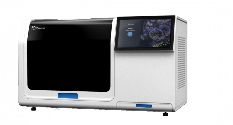
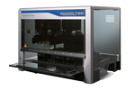
Xenium Analyzer - 10x GENOMICS
The Xenium in situ Analyzer enables the analysis of gene expression in spatial context with subcellular resolution. The results provide a detailed map of gene expression while preserving the cellular localization of hundreds of RNA targets directly within the tissue, without the need for conventional sequencing. When combined with panels, powerful visualization software, and an automated workflow, the Xenium Analyzer forms a robust platform for multiplex in situ profiling.
.png)
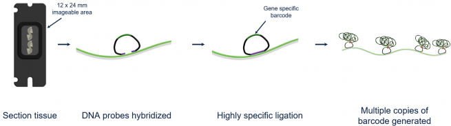
The Xenium Analyzer allows the processing of two chips, each with a 12 × 24 mm imaging area, in just two days. Its simple workflow and instrument design enable you to move quickly from sample to results.
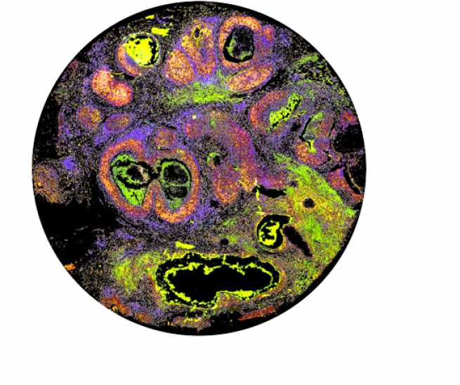
The Xenium in situ analysis uses specific probes for target transcripts capable of circularization, followed by enzymatic amplification. On the Xenium Analyzer, microscopic images of the tissue detect the position of each fluorescent probe, which is then removed. Subsequent rounds of fluorescent probe hybridization, imaging, and removal create a unique pattern of fluorescent colors at a specific site, forming a distinct optical signature that reveals the RNA identity at that location within each cell of the tissue. In the future, Xenium will also enable the detection of both RNA and protein within the same tissue section.